Adding nanoparticles to the surface of tumour cells could make them more susceptible to treatment with particular cancer drugs, according to new research at MIT. The study showed that nanoparticles tethered to the cell surface can increase the effects of forces exerted on the tumour cells by physiological fluids flowing within the body, which makes the cells much more vulnerable to attack by certain therapeutics.
Scientists have recently been exploring the physical properties of tumours and their microenvironment, with recent research showing that tumours can exploit the forces in their surroundings to enhance their survival and promote the progression of the cancer. But researchers at MIT believe that increasing the forces exerted on tumour cells can make certain therapeutics more effective in killing cells and controlling the cancer.
The study, which was led by Robert Langer, used an experimental drug known as TRAIL (a TNF-related apoptosis-inducing ligand), which exerts a cytotoxic effect on tumour cells without damaging healthy cells. TRAIL also avoids many of the debilitating effects of more commonly used therapies.
The researchers found that when used to target tumour cells, TRAIL was more successful in killing cancerous cells once they had been exposed to the shear forces generated by physiological fluids such as blood flow in the body (Nat. Commun. 8 14179). The MIT team set out to optimize the forces required for cell death, and they found that increasing the force on the cells made them more susceptible to TRAIL.
Nanoparticles strengthen forces
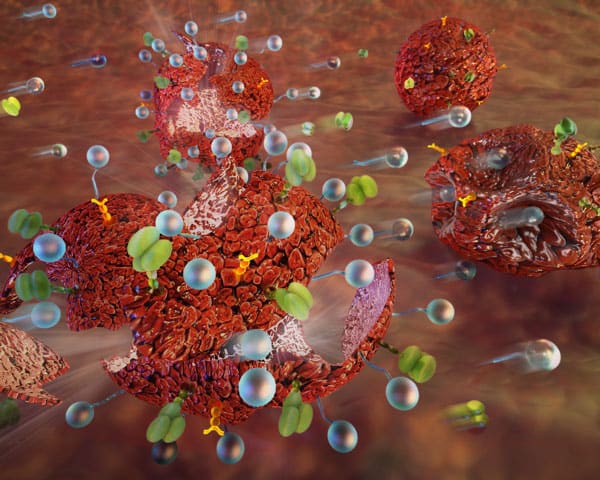
To produce these forces, Langer and his colleagues have pioneered the use of nanoparticles made from biodegradable polymers known as PLGA (poly(lactic-co-glycolic acid)). When injected into the bloodstream, the nanoparticles attach to the tumour cell surface and increase the force on the cell from the flow of physiological fluids.
The nanoparticles are coated with PEG (polyethylene glycol), which is tagged with a specific ligand that interacts with proteins found on the surface of the tumour cells. These ligands, and therefore the nanoparticles, are attached to the tumour cell like a ball tied to a string. The shear forces from the flow of physiological fluids cause the nanoparticles to bump and bash the tumour cells, causing them to become more susceptible to the effects of the therapeutics.
The MIT team found that attaching the nanoparticles to tumour cells prior to treatment with TRAIL killed metastatic tumours and reduced the progression of tumours in mice. The researchers also found that the treatment appeared to be specific to tumour cells and that healthy cells remained unaffected. Nanoparticle size and quantity were also found to have an effect on cell death. Larger particles, around one micrometre across, and a higher quantity of particles were found to have the most positive effect.
The researchers believe that the mechanism the nanoparticles induce on the tumour cells may cause the molecules surrounding the tumour cells to compress, enabling the therapeutics to interact more efficiently with receptors on the cell surface.
Langer and his research team are now exploring the possibilities of using this technique in combination with other drugs. This is a key strategy to prevent drug resistance in cancer treatment since tumours often regrow and become unaffected by drugs that had previously been effective. The MIT team is particularly interested in drug combinations that induce cytokines, which stimulate signalling chemicals that trigger an immune response to the site that helps destroy the tumour.

