By incorporating divalent cations into alginate solutions, PhD student Thomas Valentin and colleagues at Brown University in the US have produced a noncovalently crosslinked hydrogel that is responsive to multiple stimuli. The biocompatible material, derived from algae, was used to create temporary ordered structures that could be further used as templates for fabricating more complex architectures. The researchers demonstrated the versatility of this process by creating microfluidic channels, and degradable barriers were found to direct cellular migration.
As crosslinked polymeric networks with high water content, hydrogels are an obvious choice in many different areas of research. Despite containing a lot of water, they will exhibit solid-like behaviour due to strong interactions between the polymer chains known as crosslinks. However, across all fields, the use of hydrogels is limited by the lack of control over the inner architecture of these crosslinked polymer networks.
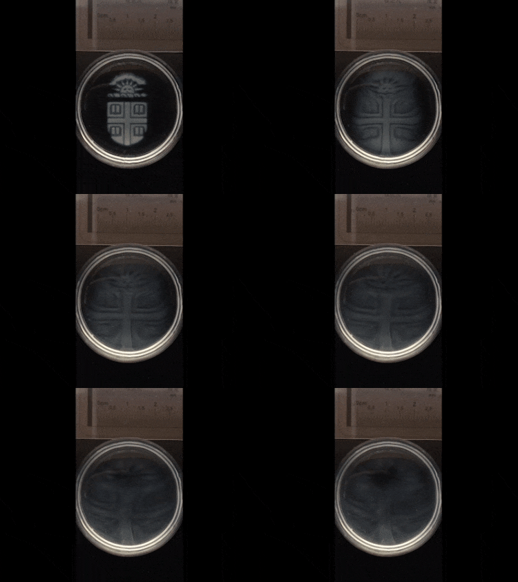
Valentin and his colleagues used a hydrogel solution composed of alginate, metal-based cationic salts and photoinitiators. Upon exposure to a laser, the light-sensitive photoinitiators enabled the metal salts to dissolve in solution, which generated free metal cations. These cations bound to the polymer and, due to their charge, facilitated ionic crosslinking between the polymer chains. Sequential layers of hydrogel can be crosslinked through this “bottom-up” technique known as photopatterning, facilitating the production of 3D structures. Having optimized the gel composition, the researchers used light-based 3D printing to fabricate stepped structures 1.7 mm tall with a vertical resolution of 250 μm.
In contrast to conventional hydrogel crosslinking routes based on permanent covalent bonds between polymers, the ionic crosslinking is entirely reversible, enabling on demand, temporally rapid, degradation of the hydrogel material. This charge interaction is entirely reversible through chelation, where ions bind to metals. The addition of a solution containing chelating agents will dissociate the metal cations bound to polymers, thus disrupting the crosslinking and hydrogel network.
Following this new method, the alginate gels successfully templated a secondary material; agarose, a hydrogel derived from seaweed. Agarose gelled around the features already patterned within the alginate gel. Addition of chelating agents then degraded the alginate gel templates. Unaffected by this process, the agarose gel remained intact, with hollow channels where the alginate gel once was. Senior author Ian Wong describes the patterning and degradation process as being “a bit like Lego bricks. We can attach polymers together to build 3-D structures, and then gently detach them again under biocompatible conditions.”
Utilising the biocompatibility of the material, controlled patterning and degradation of the alginate hydrogel revealed new cell migration behaviour in mammary cells. Structures patterned in the alginate hydrogels spatially limited the growth of the cells within the network to certain regions. Subsequent degradation of the alginate presented unoccupied regions to the cells, which they moved into. Collective cell behaviour revealed that migration of the frontmost cells would act to straighten out the initial geometrical shapes where the cells had initially been patterned. The cells moved to occupy all available regions within two days, showing no compromise in cell viability throughout the patterning and degradation processes.
Future work is planned to improve the spatial resolution as well as control of the mechanical properties and degradation kinetics. The development of this 3D technique for patterning gels also opens the door for other ionically crosslinked gels to be patterned in this way, revolutionizing many different areas of research.
Full details of the work are reported in Lab on a Chip.


