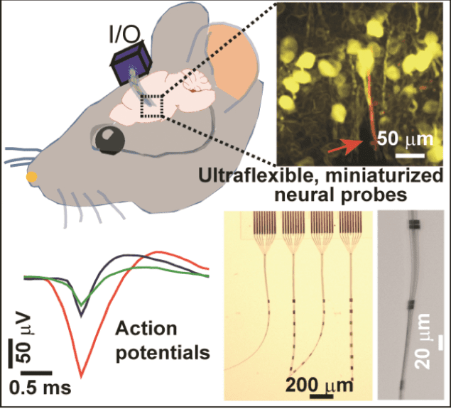
Nanofabricated ultraflexible electrode arrays that have a cross-sectional area that is much smaller than conventional carbon fibre electrodes could be used to detect action potentials from individual neurons without damaging brain tissue. The devices could allow for high-density electrical recoding of neural activity in the living brain as well as being useful in the field of brain-machine interfacing.
Implanted neural probes are currently the only way to monitor the fast electrophysiological activity of individual neurons. Such probes have also been successfully employed to treat a number of neurological diseases, such as Parkinson’s. And they can be used to establish direct communication between the brain and a man-made interface too.
The problem is that these probes are typically much larger in size than neurons and capillaries (they usually have a cross-sectional area of 103 μm2) and can thus cause significant damage to brain tissue when implanted.
A hundred times smaller cross-sectional area
A team of researchers led by Chong Xie of the University of Texas at Austin has now developed new ultraflexible intracortical probes that are 10 times smaller than conventional carbon fibre electrodes and which have a cross-sectional area that is a hundred times smaller. “This advance is, in our opinion a crucial first step towards high density, full-coverage mapping of neural activity in a sizeable brain region,” says team member and lead author of this study Xiaoling Wei. “It could also prove very useful in the field of brain-machine interfacing.”
The researchers used state-of-the-art electron-beam lithography and photolithography techniques to fabricate nanoelectronic thread (NET-e) probes containing densely packed electrode arrays. They made the devices on 100 mm silicon wafers containing a sacrificial layer of thin nickel. “After releasing the sacrificial layer, we were able to harvest substrate-less small-sized probes with a multilayer layout that markedly reduces their cross-sectional area to the subcellular range,” explains Wei. “One such NTE-e hosted a linear array of electrodes with a cross-sectional profile of 0.8μm × 8 μm, which is the smallest among all reported to our knowledge.”
No damage to brain tissue
One of the probes was designed to function as a multiple tetrode, hosting groups of four closely-spaced electrodes every 100 μm along the thread, he adds. “It was fabricated so that its electrode spacing was smaller than its detection range to allow for ‘spatial oversampling’, which improves spatial resolution of action potential signal recording.”
The researchers tested out their device on a mouse model and found that the electrodes can record action and local field potentials of neurons with high signal-to-noise ratio. “Compared to conventional neural probes, the much-reduced dimensions of our NET-e probes allow us to implant the devices at previously unattainable high densities without damage to brain tissue,” Wei tells nanotechweb.org. “In this work, we could implant the probes to a depth of between 500 and 700 μm in the somatosensory cortex. We also demonstrated implantation at inter-probe distances of 60 μm distances, again without eliciting neuronal death.”
The team, reporting its work in Advanced Science https://doi.org/10.1002/advs.201700625, is now planning to extend its tests to primates. “We also plan to collaborate with other professionals to broaden the use of our probes to other fields, such as monitoring neuronal disease, and in fundamental studies.”


