Bone is certainly an impressive natural material because it is both stiff (it endures load without deforming much) and tough (it endures load and deformation without breaking). Stiffness and toughness are rarely found together in materials. The strength of bone comes from two main components: the mineral calcium phosphate (of the apatite variety), and the protein collagen.
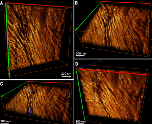
The way in which bone collagen and mineral organize themselves has proved difficult to understand, however, because the way they assemble is complex at every length scale. Scanning transmission electron microscopy tomography experiments by researchers led by Roland Kröger of the University of York and Molly Stevens of Imperial College London, both in the UK,have now revealed, among other things, that bone mineral is hierarchically organized from the nanoscale upwards. It is composed of curved needle-like crystallites that merge into twisted plates that form larger aggregates, thus creating a bridge between adjacent collagen fibrils.
The findings could be important for a variety of fields, including osteology and archeology. They will also provide valuable insight into skeletal diseases, growth and development, and might even help in the design of novel biomimetic materials.
Evaluating the mineral phase of bone in 3D
Kröger and Stevens set out to evaluate the mineral phase of bone in 3D. They used electron tomography – that means collecting images at many sequential angles or “tilts” – to reconstruct 3D images of their samples.
One of their observations, for example, was the slight curvature of crystallites across their lengths. Another was stacks of parallel twisted crystallites around irregular voids in the oblique direction. Finally, they saw tight packing of hexagonal crystals into rosette shapes in perpendicular projections. “We developed a model that puts all these observations together that shows that crystals are curved, hierarchical and splaying to form a continuous cross-fibrillar network,” explain Kröger and Stevens.
Fractal-like helical motifs
“The twisting crystals in bone assemble with twisted collagen chains and form fibrils that are twisted themselves”, they add. “As known from earlier studies, the fibrils twist into concentric layers that gently twist themselves around tiny capillaries in bone, which in turn twist around the bone’s long axis, which is often curved into a helix (like a rib, or a collar bone).”
The researchers say that the helical twist is self-similar, or fractal-like. Fractal-like helical motifs are ubiquitous in nature (think of ferns or cauliflower). “This type of organization might have evolved over millions of years to optimize bone structure and function,” lead author of this study Natalie Reznikov tells nanotechweb.org.
Yong Mao of the University of Nottingham in the UK agrees: “This is certainly an interesting study, contributing to the understanding of bone structure,” he comments. “I think researchers are increasingly appreciating the fractal/hierarchical nature of the world, which generally offers an evolutionary advantage. These finer structures are becoming more accessible with the development of additive manufacturing (or 3D printing). I expect this to be an exciting field of development in the future.
The research is detailed in Science 10.1126/science.aao2189.
For more on hierarchical nanostructures visit the Nanotechnology focus collection.


